|
A cardiac stress PET scan, the definition !
What is a cardiac PET scan? A PET scan of the heart is a noninvasive nuclear imaging test. It uses radioactive tracers (called radionuclides) to produce pictures of your heart. Doctors use cardiac PET scans to diagnose coronary artery disease (CAD) and damage due to a heart attack. PET scans can show healthy and damaged heart muscle. Doctors also use PET scans to help find out if you will benefit from a percutaneous coronary intervention (PCI) such as angioplasty and stenting, coronary artery bypass surgery (CABG) or another procedure. Quick facts PET scans use radioactive material called tracers. Tracers mix with your blood and are taken up by your heart muscle. A special “gamma” detector that circles the chest picks up signals from the tracer. A computer converts the signals into pictures of your heart at work. A PET scan shows if your heart is getting enough blood or if blood flow is reduced because of narrowed arteries. It also shows dead cells (scars) from a prior heart attack. A PET scan can help in determining if you’ll benefit from a cardiac procedure (PCI) or surgery to restore blood flow. The tracers used for PET scans can help identify injured but still living (viable) heart muscle that might be saved if blood flow is restored. Why do people have cardiac PET scans? A PET scan is a very accurate way to diagnose coronary artery disease and detect areas of low blood flow in the heart. PET can also identify dead tissue and injured tissue that’s still living and functioning. If the tissue is viable, you may benefit from a PCI or coronary artery bypass surgery. How does a PET scan work? A radioactive tracer is injected into your bloodstream. The tracers used for PET are mostly natural body compounds such as glucose, water or ammonia, which are labeled or “tagged” with a small amount of radioactive material. Inside your body the radioactive tracer produces a type of energy called a gamma ray. Gamma rays are detected by a gamma detector and are used to produce a series of clear images of your heart. Images of thin slices made all the way through the heart can be produced from all different directions and angles. Computer graphics can be used to create a 3-dimensional image of your heart from the thin-slice images. Your doctor will be able to tell if your heart muscle is functioning or (if the heart muscle isn’t working) if your heart muscle is still alive by how well it takes up and uses the different tracers. The doctor will determine this by examining the images to find where the tracer is. Viable heart tissue will take in more of the tracer than tissue that’s no longer viable. Different colors or degrees of brightness on the PET scan show different levels of tissue function. What are the risks of cardiac PET? Cardiac PET is safe for most people. The amount of radiation is small, and your body will get rid of it through your kidneys within about 24 hours. If you’re pregnant or think you might be pregnant, or if you’re a nursing mother, tell your doctor before you have this test. It could harm your baby. How do I prepare for cardiac PET? Tell your doctor about any medicines you take, including over-the-counter medicines, herbs and vitamins. Your doctor may ask you not to take them before the test. Don’t stop taking your medicine until your doctor tells you to. If you have diabetes and take insulin, talk with your doctor about how much insulin you should take before the scan and what you should eat. Your blood sugar levels will be monitored during the test. Test results are not always accurate in people with diabetes. Your doctor may also ask you to avoid certain foods and drinks, such as caffeine-containing drinks or alcohol, for 24 hours before your test. Don’t eat, and drink only water for 4 to 6 hours before your test. Wear comfortable, loose-fitting clothing, and don’t wear jewelry or other metal objects. What happens during cardiac PET? A doctor and a nuclear medicine technologist usually perform the scan in a hospital or at a PET center using special equipment. The technician will place small metal disks (electrodes) on your chest, arms and legs. The disks have wires that hook to a machine that records your electrocardiogram (ECG or EKG). The ECG keeps track of your heartbeat during the test and can signal the computer when to take a scan. The technician will put an intravenous line (IV) in your arm. The tracer will be injected through the IV line. You’ll lie on a flat table that’s connected to the PET scanner and a computer. The table will slide into the scanner, which is shaped like a giant doughnut. Within the PET scanner, detectors record the radioactive patterns of the tracer in your heart. The information is transformed into images on a computer screen. Several scans are done over time to provide pictures of thin slices of your entire heart from all angles. It’s very important to hold completely still with your arms above your head while each scan is being done. Doctors will take a baseline picture of your heart before the tracer is injected. This takes about 15 to 30 minutes. Next, the tracer will be injected and your heart will be scanned again. If you will have a nuclear chemical stress test (also called a pharmacologic stress test), you’ll get a medicine that increases the blood flow in your heart, similar to what happens during exercise. These medicines may include adenosine, dipyridamole (Persantine) or dobutamine. The doctor will examine how well your heart takes up the tracer before and after receiving the medicine. If you have severe coronary artery disease, some areas of your heart may not get enough blood during a stress so the tracer won’t show up in those areas. The test takes between 1 and 3 hours. What happens after my PET scan? You can usually go back to your normal activities right away. Drink plenty of water for the next 24 hours to flush the radioactive material from your body. Make an appointment with your doctor to discuss the results of the test and next steps. How can I learn more about cardiac PET? Talk with your doctor. Here are some good questions to ask: Why are you doing this test rather than a different one? What do I need to do to get ready for this test? When will I get my test results? Will I need to have more tests after this?
1 Comment
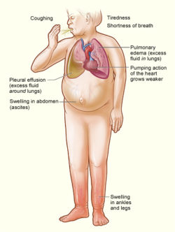 When it comes to congestive heart failure, we get a lot of questions from our patients regarding symptoms and treatments options. In fact, we have a new Transitional Heart Failure Clinic opening specifically to help treat those patients. But, what is congestive heart failure and how can it be treated?
|
AuthorWrite something about yourself. No need to be fancy, just an overview. ArchivesCategories |
 RSS Feed
